45 microscope images with labels
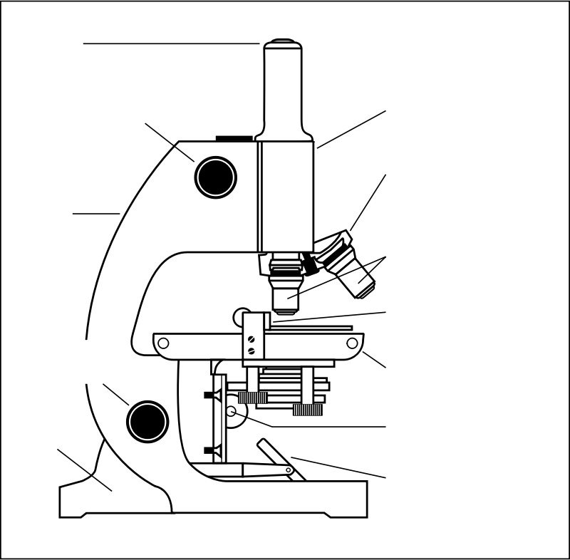
Parts of the Microscope with Labeling (also Free Printouts) Microscopes are specially created to magnify the image of the subject being studied. This exercise is created to be used in homes and schools. the microscope layout, including the blank and answered versions are available as pdf downloads. Click to Download : Label the Parts of the Microscope (A4) PDF print version. › blog › postsLyft's Commitment to Climate Action - Lyft Blog A lot of voters agree with us. Early support for the measure is strong. What started with good policy created by a diverse group of organizations — including the Natural Resources Defense Council, the American Lung Association, California State Firefighters, the Coalition for Clean Air, the State Association of Electrical Workers – IBEW, the San Francisco Bay Area Planning and Urban ...
Avery Self-Adhesive Removable Labels, 0.75 x 1 Inches, White, … Apr 18, 2005 · Buy Avery Self-Adhesive Removable Labels, 0.75 x 1 Inches, White, 1000 per Pack (05428): ... IMAGES ; Avery Self-Adhesive Removable Labels, 0.75 x 1 Inches, White, 1000 per Pack (05428) ... fits 1 x 3 inch microscope slides which is what I wanted to use it for. Read more. Report abuse. Abha.
Microscope images with labels
PPIC Statewide Survey: Californians and Their Government Oct 27, 2022 · Key Findings. California voters have now received their mail ballots, and the November 8 general election has entered its final stage. Amid rising prices and economic uncertainty—as well as deep partisan divisions over social and political issues—Californians are processing a great deal of information to help them choose state constitutional officers and … Parts of a microscope with functions and labeled diagram - Microbe Notes Parts of a microscope with functions and labeled diagram September 17, 2022 by Faith Mokobi Having been constructed in the 16th Century, Microscopes have revolutionalized science with their ability to magnify small objects such as microbial cells, producing images with definitive structures that are identifiable and characterizable. Lyft's Commitment to Climate Action - Lyft Blog A lot of voters agree with us. Early support for the measure is strong. What started with good policy created by a diverse group of organizations — including the Natural Resources Defense Council, the American Lung Association, California State Firefighters, the Coalition for Clean Air, the State Association of Electrical Workers – IBEW, the San Francisco Bay Area Planning and …
Microscope images with labels. › articles › s41551/020/00682-wData-efficient and weakly supervised computational pathology ... Mar 01, 2021 · A data-efficient and interpretable deep-learning method for the multi-class classification of whole-slide images that relies only on slide-level labels is applied to the detection of lymph node ... › publication › ppic-statewide-surveyPPIC Statewide Survey: Californians and Their Government Oct 27, 2022 · Key Findings. California voters have now received their mail ballots, and the November 8 general election has entered its final stage. Amid rising prices and economic uncertainty—as well as deep partisan divisions over social and political issues—Californians are processing a great deal of information to help them choose state constitutional officers and state legislators and to make ... AX / AX R | Confocal Microscopes | Nikon Microscope Products Nikon’s newest confocal microscope with unparalleled resolution, speed, sensitivity and field of view, with AI-based tools for simplifying acquisition and analysis. ... The AX/AX R’s all new DUX-VB detector custom-tunes emission bandwidths to a library of labels and probes, and provides the freedom to fine-tune emission bands to minimize ... Data-efficient and weakly supervised computational pathology ... - Nature Mar 01, 2021 · A data-efficient and interpretable deep-learning method for the multi-class classification of whole-slide images that relies only on slide-level labels is applied to the detection of lymph node ...
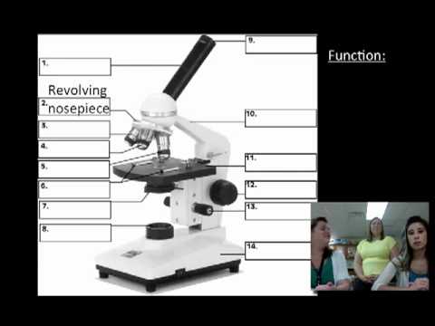
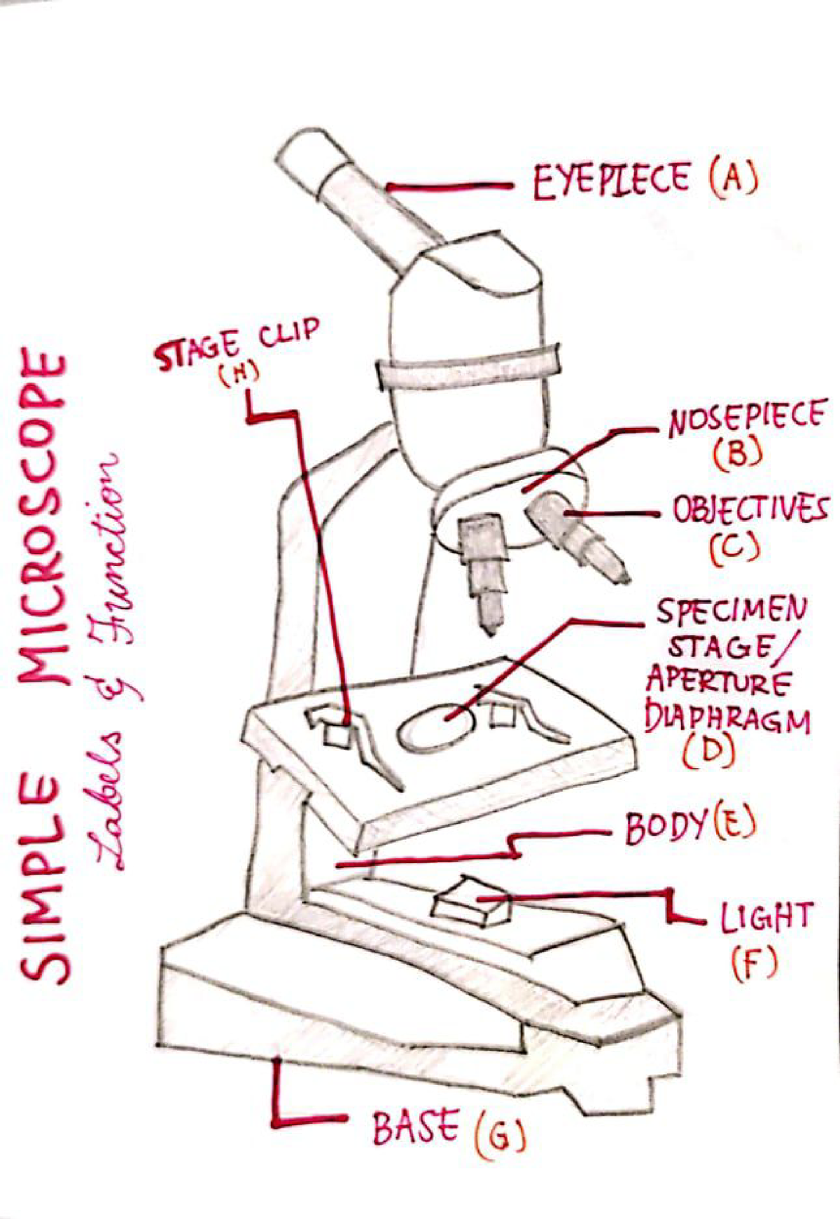
Microscope Types (with labeled diagrams) and Functions The working principle of a simple microscope is that when a lens is held close to the eye, a virtual, magnified and erect image of a specimen is formed at the least possible distance from which a human eye can discern objects clearly. Simple microscope labeled diagram Simple microscope functions It is used in industrial applications like: Microscope Labeling - The Biology Corner The google slides shown below have the same microscope image with the labels for students to copy. I often spend the first day walking students through the steps and having them look at a single slide as we do the steps. Students are often very enthusiastic about using microscopes and will try to start with the high power objective. Microscope Images Labeled | Virtual Anatomy Lab VAL - ncccval Body cavities, planes, and regions. Body Images Labeled. Body Images Unlabeled. Histology. Epithelium Images Labeled. Epithelium Images Unlabeled. Connective Tissue Images Labeled. Connective Tissue Images Unlabeled. Microscope. Compound Microscope Parts - Labeled Diagram and their Functions The eyepiece (or ocular lens) is the lens part at the top of a microscope that the viewer looks through. The standard eyepiece has a magnification of 10x. You may exchange with an optional eyepiece ranging from 5x - 30x. [In this figure] The structure inside an eyepiece. The current design of the eyepiece is no longer a single convex lens.
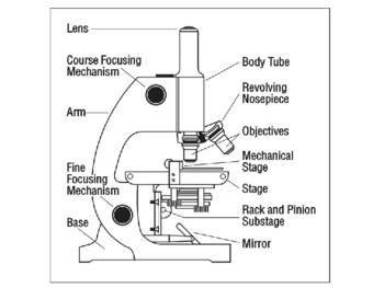
Amazon.com: microscope slide labels Microscope Slide Label SLS-15, Standard, 1000/PK. $33.00 $ 33. 00. Get it Wed, Oct 19 - Mon, Oct 24. $8.00 shipping. Only 10 left in stock - order soon. Small Business. Small Business. Shop products from small business brands sold in Amazon's store. Discover more about the small businesses partnering with Amazon and Amazon's commitment to ... Parts of Stereo Microscope (Dissecting microscope) – labeled … A Stereo microscope is like a powerful magnifying glass, good for thick and solid specimens for observing the surface textures with 3D vision. ... images into a three-dimensional vision. [In this image] The concept of 3-D vision is the same in both 3-D movies and stereo microscopes. The key is presenting two images with a slightly different ... Microscope Parts and Functions Body tube (Head): The body tube connects the eyepiece to the objective lenses. Arm: The arm connects the body tube to the base of the microscope. Coarse adjustment: Brings the specimen into general focus. Fine adjustment: Fine tunes the focus and increases the detail of the specimen. Nosepiece: A rotating turret that houses the objective lenses. Laser Scanning Confocal Microscopy | Nikon’s MicroscopyU In addition, necessary low concentrations of fluorescent labels usually produce a weak signal having poor contrast. Another concern is that long exposure to intense low-wavelength illumination often limits cell and tissue viability and, consequently, the length of experiments. ... A confocal microscope that captures images with a 25 mm field of ...
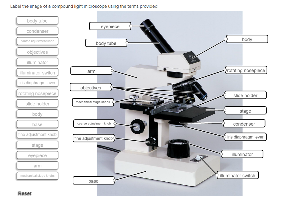
Solved Label the image of a compound light microscope using ...
Compound Microscope - Diagram (Parts labelled), Principle and Uses See: Labeled Diagram showing differences between compound and simple microscope parts Structural Components The three structural components include 1. Head This is the upper part of the microscope that houses the optical parts 2. Arm This part connects the head with the base and provides stability to the microscope.

Labeling the Parts of the Microscope | Microscope activity ...
Skin Images Labeled | Virtual Anatomy Lab VAL - ncccval Body cavities, planes, and regions. Body Images Labeled. Body Images Unlabeled. Histology. Epithelium Images Labeled. Epithelium Images Unlabeled. Connective Tissue Images Labeled. Connective Tissue Images Unlabeled. Microscope.

Microscope With Labels free vector | Download it now!
› Hayve-Microscope-MagnificationAmazon.com : Hayve 7" LCD Digital Microscope, 1200X ... Jul 25, 2022 · LCD Digital Microscope, ANNLOV 4.3 Inch Handheld USB Microscope 50X-1000X Magnification Coin Microscope with 32GB TF Card, 8 Adjustable LED, PC View, Microscope for Adults/Kids Add to Cart Customer Rating

microscope drawing with label - Clip Art Library
400+ Free Microscope & Bacteria Images - Pixabay 413 Free images of Microscope Related Images: bacteria science laboratory research scientist biology lab chemistry microbiology Find your perfect microscope image. Free pictures to download and use in your next project. Next page ›

Microscope Diagram Labeled, Unlabeled and Blank | Parts of a ...
› products › conAX / AX R | Confocal Microscopes | Nikon Microscope Products ... The AX R’s high speed resonant scanning, which decreases the illumination time by more than 20x typical confocal scanning times, greatly reduces biases caused by merely acquiring images. Reducing the acquisition time also allows for extremely high-speed imaging (up to 720 fps @ 2048 x 16).
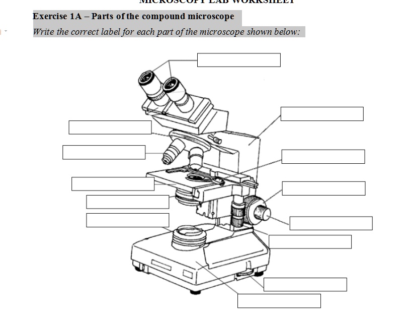
SOLVED: Exercise 1A Parts ofthe compound microscope Write the ...
Microscope Labeled Pictures, Images and Stock Photos Browse 49 microscope labeled stock photos and images available, or start a new search to explore more stock photos and images. Newest results Fluorescent Imaging immunofluorescence of cancer cells growing... Microscope diagram vector illustration. Labeled zoom instrument... Microscope diagram vector illustration.
Microscope With Labels - Free Transparent PNG Download - PNGkey
Mitosis Images Labeled | Virtual Anatomy Lab VAL - ncccval Endocrine Rabbit Dissection Unlabeled. Cardiovascular. Cardiovascular Histology Labeled. Cardiovascular Histology Unlabeled. Cardiovascular Models Labeled. Cardiovascular Models Unlabeled. Cardiovascular Sheep Heart Dissect-L. Cardiovascular Sheep Heart Disect-U. Cardiovascular Cat Dissection Labeled.

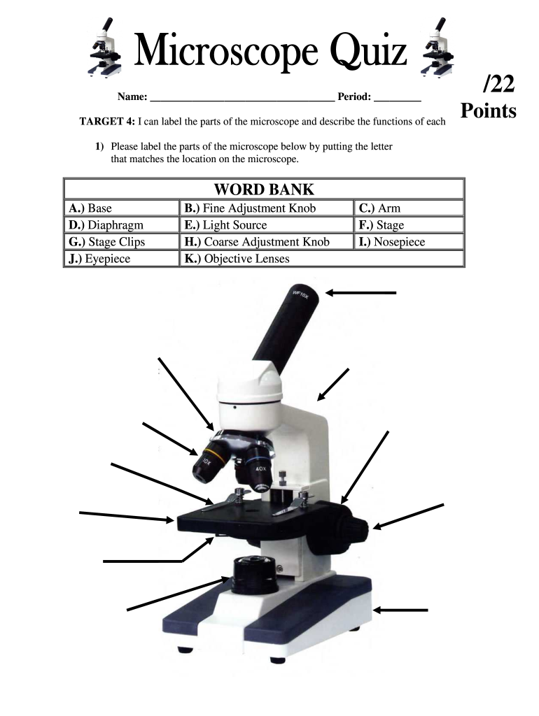
Label a Microscope Worksheet
rsscience.com › stereo-microscopeParts of Stereo Microscope (Dissecting microscope) – labeled ... Unlike a compound microscope that offers a flat image, stereo microscopes give the viewer a 3-dimensional image that you can see the texture of a larger specimen. [In this image] Examples of Stereo & Dissecting microscopes. Major microscope brands (Zeiss, Olympus, Nikon, Amscope, Omano, Leica …) all produce stereomicroscopes.

Label the numbered parts of the microscope - ppt download
Label the microscope — Science Learning Hub All microscopes share features in common. In this interactive, you can label the different parts of a microscope. Use this with the Microscope parts activity to help students identify and label the main parts of a microscope and then describe their functions. Drag and drop the text labels onto the microscope diagram.
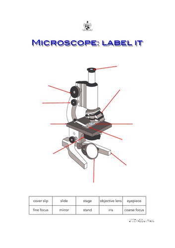
Microscope: label it | Teaching Resources
Microscope Images Unlabeled | Virtual Anatomy Lab VAL - ncccval Body cavities, planes, and regions. Body Images Labeled. Body Images Unlabeled. Histology. Epithelium Images Labeled. Epithelium Images Unlabeled. Connective Tissue Images Labeled. Connective Tissue Images Unlabeled. Microscope.

Compound Microscope Parts – Labeled Diagram and their ...
How to Use the Microscope - The Biology Corner Light Microscope - the models found in most schools, use compound lenses to magnify objects. The lenses bend or refract light to make the object beneath them appear closer. ... All drawings should include clear and proper labels (and be large enough to view details). Drawings should be labeled with the specimen name and magnification ...
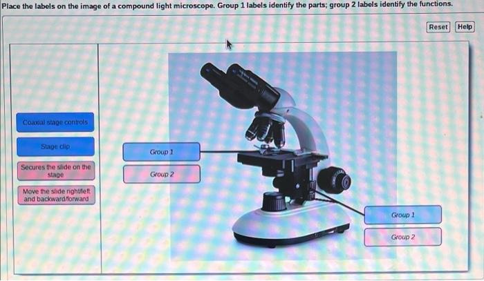
Solved Place the labels on the image of a compound light ...
Super-resolution microscopy - Wikipedia Super-resolution microscopy is a series of techniques in optical microscopy that allow such images to have resolutions higher than those imposed by the diffraction limit, which is due to the diffraction of light. Super-resolution imaging techniques rely on the near-field (photon-tunneling microscopy as well as those that utilize the Pendry Superlens and near field scanning optical …
microscope with labels - Openclipart
Explanation and Labelled Images - New York Microscope Company Another use of fluorescence imaging is Fluorescence Speckle Microscopy. It is a technology that uses fluorescence labeled macromolecular assemblies such as cytoskeletal protein to study movement and turnover rates. Fluorescence microscopy staining also is helpful in the field of mineralogical applications. It is routinely used for the study of ...

Biology Lab Quiz #2 (Labeling a Microscope) Diagram | Quizlet
Compound Microscope Parts, Functions, and Labeled Diagram Compound Microscope Definitions for Labels. Eyepiece (ocular lens) with or without Pointer: The part that is looked through at the top of the compound microscope. Eyepieces typically have a magnification between 5x & 30x. Monocular or Binocular Head: Structural support that holds & connects the eyepieces to the objective lenses.
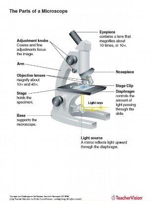
Microscope Parts and Functions
Microscope Labels Photos - Free & Royalty-Free Stock Photos from Dreamstime Download all free or royalty-free photos and images. Use them in commercial designs under lifetime, perpetual & worldwide rights. Dreamstime is the world`s largest stock photography community.
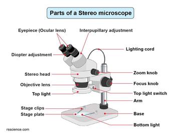
Parts of Stereo Microscope (Dissecting microscope) – labeled ...
Microscope Labeling Game - PurposeGames.com About this Quiz. This is an online quiz called Microscope Labeling Game. There is a printable worksheet available for download here so you can take the quiz with pen and paper. This quiz has tags. Click on the tags below to find other quizzes on the same subject. Science.

Microscope Diagram Labeled, Unlabeled and Blank | Parts of a ...
Amazon.com : Hayve 7" LCD Digital Microscope, 1200X … Jul 25, 2022 · LCD Digital Microscope, 1000X Coin Microscope Gift with a Guidebook, Microscope for Adults/Kids Video Microscope with 32GB TF Card, Windows Compatible 7 inch LCD Digital Microscope ANNLOV 1-1200X USB Maginfication 12MP Handheld Electronic Coin Microscope Camera with 8 Adjustable LED Lights for Adults PCB Soldering Kids Outside Use

Meiji MT6500 Series PCM NIOSH 7400 Asbestos Microscope
› worksheets › microscope_useHow to Use the Microscope - The Biology Corner Stereoscope - this microscope allows for binocular (two eyes) viewing of larger specimens. Scanning Electron Microscope - allow scientists to view a universe too small to be seen with a light microscope. SEMs do not use light waves; they use electrons (negatively charged electrical particles) to magnify objects up to two million times.

What is a Compound Microscope? | Microscope World Blog
Labeling the Parts of the Microscope | Microscope World Resources Labeling the Parts of the Microscope This activity has been designed for use in homes and schools. Each microscope layout (both blank and the version with answers) are available as PDF downloads. You can view a more in-depth review of each part of the microscope here. Download the Label the Parts of the Microscope PDF printable version here.
Microscope with labels picture
Lyft's Commitment to Climate Action - Lyft Blog A lot of voters agree with us. Early support for the measure is strong. What started with good policy created by a diverse group of organizations — including the Natural Resources Defense Council, the American Lung Association, California State Firefighters, the Coalition for Clean Air, the State Association of Electrical Workers – IBEW, the San Francisco Bay Area Planning and …

Solved Microscope parts/labeling 9 Label the image of a ...
Parts of a microscope with functions and labeled diagram - Microbe Notes Parts of a microscope with functions and labeled diagram September 17, 2022 by Faith Mokobi Having been constructed in the 16th Century, Microscopes have revolutionalized science with their ability to magnify small objects such as microbial cells, producing images with definitive structures that are identifiable and characterizable.
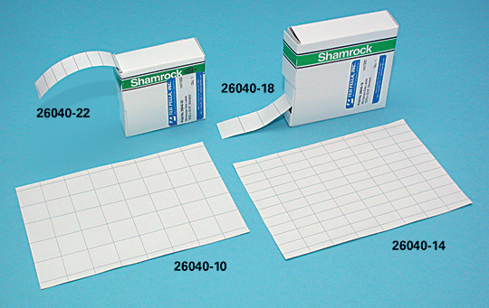
Microscope Slide Labels
PPIC Statewide Survey: Californians and Their Government Oct 27, 2022 · Key Findings. California voters have now received their mail ballots, and the November 8 general election has entered its final stage. Amid rising prices and economic uncertainty—as well as deep partisan divisions over social and political issues—Californians are processing a great deal of information to help them choose state constitutional officers and …
Microscope- Simple-AND Compound-WITH- Label - BS in Education ...

label microscope diagram | Charts | Microscope, Anatomy bones ...

Label The Microscope Parts! Diagram | Quizlet

Parts of the Microscope with Labeling (also Free Printouts ...
Microscope Fill In The Blank - Fill Online, Printable ...

FUNRUI Kids Microscope, 450x, 200x, 100x Magnification Children Science Microscope Kit with LED Lights Includes Accessory Toy Set for Beginners Early ...

Below is a photo of a compound light microscope with labels ...

This is a common compound microscope. Label its parts from A ...

Educational / Hobby Microscope (BE211A Eco-Bino-LED)
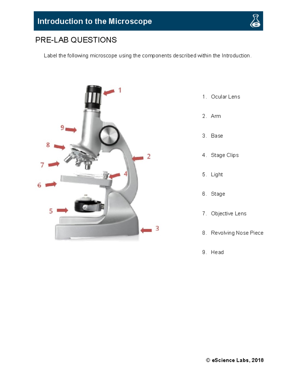
Introduction to the microscope - PRE-LAB QUESTIONS Label the ...

Parts of a Microscope Labeling Activity

The Microscope

Photo Compound microscope with labels Image #3850568
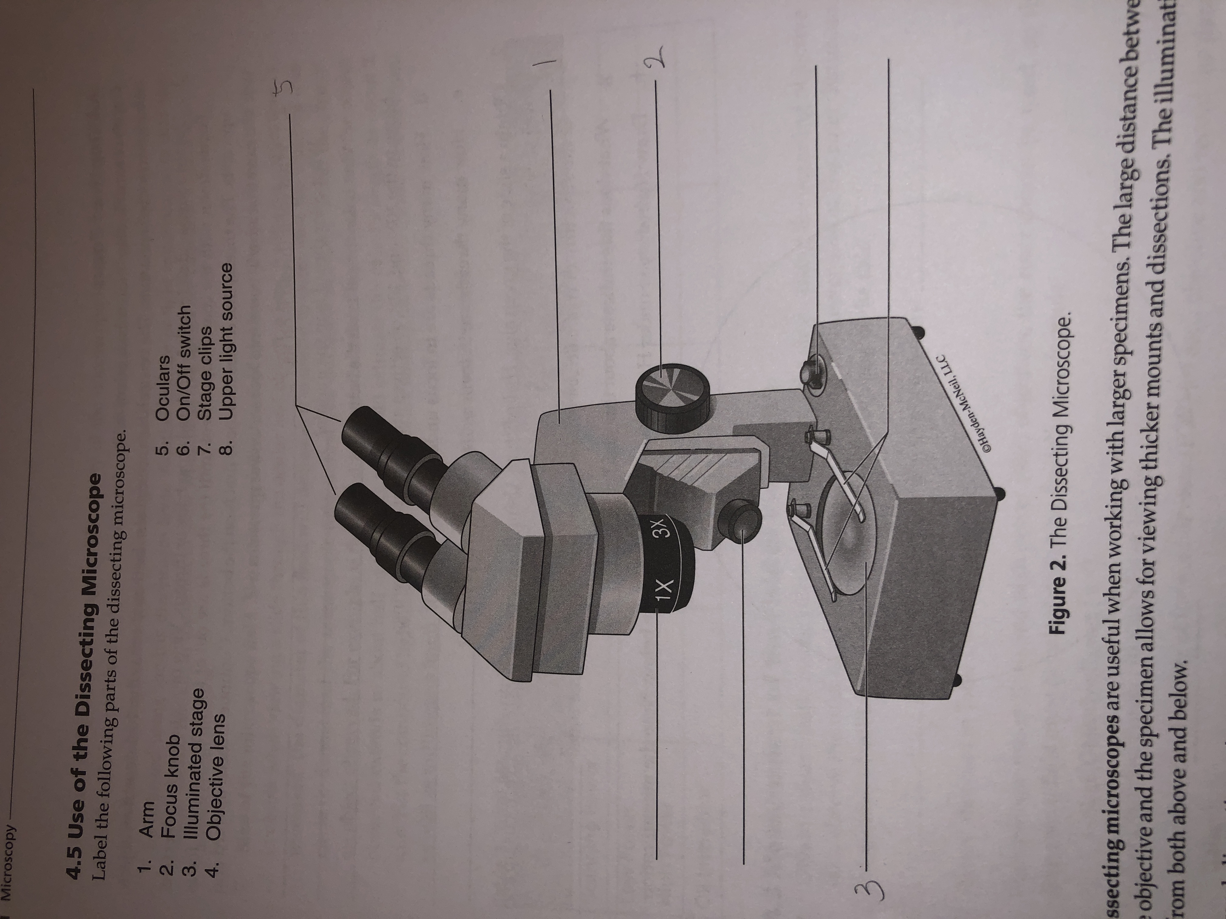
Answered: Microscopy 4.5 Use of the Dissecting… | bartleby
Compound Microscope Parts, Functions, and Labeled Diagram ...
The Parts of a Microscope (Labeled) Printable Printable (6th ...

Microscope With Labels clip art | Microscope parts, Science ...

Microscope Labeling Diagram | Quizlet

Label-microscope.docx - Label parts of the Microscope ...
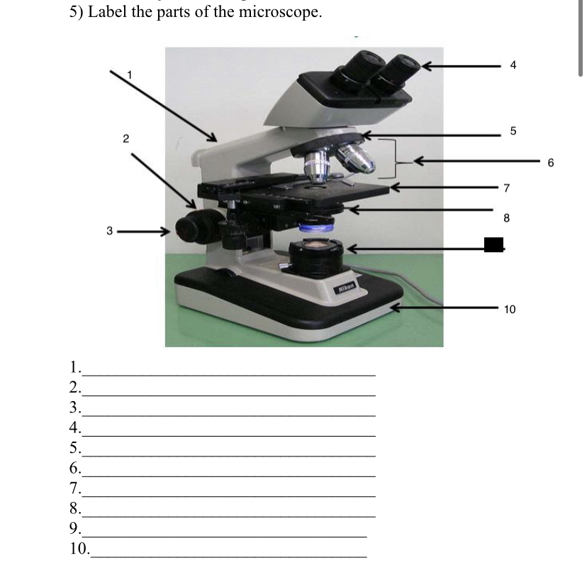
Answered: 5) Label the parts of the microscope. 1… | bartleby

Microscope Labeling #1 Diagram | Quizlet

Biology Lab Microscope Labeling Diagram | Quizlet

This is a common compound microscope. Label its parts from A ...
Post a Comment for "45 microscope images with labels"