43 cell membrane diagram with labels
A Well-labelled Diagram Of Animal Cell With Explanation Well-Labelled Diagram of Animal Cell The Cell Organelles are membrane-bound, present within the cells. There are various organelles present within the cell and are classified into three categories based on the presence or absence of membrane. Listed below are the Cell Organelles of an animal cell along with their functions. Cellular Diagram Respiration Label The Search: Cellular Respiration Label The Diagram. The Krebs cycle uses the two molecules of pyruvic acid formed in glycolysis and the performance of high-energy molecules of NADH and flavin adenine dinucleotide (FADH2), as Part 3: Aerobic Respiration The energy present in the form of ATP can then be utilized to drive various intra-cellular physiological processes like the transport of molecules ...
Label Cell Parts | Plant & Animal Cell Activity | StoryboardThat Click "Start Assignment". Find diagrams of a plant and an animal cell in the Science tab. Using arrows and Textables, label each part of the cell and describe its function. Color the text boxes to group them into organelles found in only animal cells, organelles found in only plant cells, and organelles found in both cell types.
Cell membrane diagram with labels
Diagram of a cell membrane with labels - NIST Diagram of a cell membrane with labels | NIST IMAGES Diagram of a cell membrane with labels Appears In Biology in Reflectometry Essential Biological Functions Immune response, Cell metabolism, Neurotransmission, Photosynthesis, Cell adherence, Cell growth and differentiation Potential Commercial Applications Structure of Membrane in Cells (With Diagram) - Biology Discussion Cell walls are formed through apposition, i.e., the wall material is deposited by the protoplast on the plasma membrane. The first cell wall (Primary wall) is formed during the cell growth phase. When the cell elongation is stopped the secondary cell wall, formation starts (Fig. 2.13). Cell Membrane (Plasma Membrane) - Genome.gov Definition. …. The cell membrane, also called the plasma membrane, is found in all cells and separates the interior of the cell from the outside environment. The cell membrane consists of a lipid bilayer that is semipermeable. The cell membrane regulates the transport of materials entering and exiting the cell.
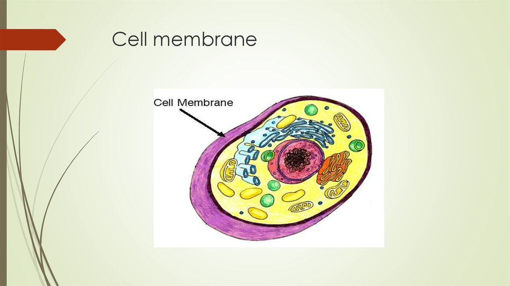
Cell membrane diagram with labels. Cell The Diagram Plant Label Which label belongs in the area marked Y? - 4883586 the cell therapy supply chainbioprocess international Plant Cell Illustration Plant Cell Illustration. diagram of plant and animal cell with labels Vacuole: A membrane-bound, fluid-filled sac inside plant and animal cells The plant parts labelling application is a simple to use science ... Cell Organelles- Definition, Structure, Functions, Diagram Cilia are hair-like projections that have a 9+2 arrangement of microtubules with a radial pattern of 9 outer microtubule doublet that surrounds two singlet microtubules. This arrangement is attached to the bottom with a basal body. Flagella is a filamentous organelle, the structure of which, is different in prokaryotes and eukaryotes. Cell Membrane - The Definitive Guide | Biology Dictionary The cell membrane, also known as the plasma membrane, is a double layer of lipids and proteins that surrounds a cell. It separates the cytoplasm (the contents of the cell) from the external environment. It is a feature of all cells, both prokaryotic and eukaryotic. a 3D diagram of the cell membrane Function of the Cell Membrane Animal Cell Diagram with Label and Explanation: Cell ... - Collegedunia Cell Membrane The cell membrane is the thin semi-permeable layer surrounding the cell; its main functionality is to protect the cell. It has hair-like structures cilia and flagella on it. The cell membrane controls the entry and exit of nutrients into the animal cell. Cytoplasm
Cell membrane - Wikipedia The cell membrane (also known as the plasma membrane (PM) or cytoplasmic membrane, and historically referred to as the plasmalemma) is a biological membrane that separates the interior of all cells from the outside environment (the extracellular space) and protects the cell from its environment. The cell membrane consists of a lipid bilayer, made up of two layers of phospholipids with ... Cell Membrane Functions, Structure and Diagram - Jotscroll Cell Membrane Diagram. Structure of a Cell membrane (Plasma Membrane) Photo Credit: Plasma Membrane Structure. The cell membrane is extremely thin measuring about 7.5-10 nm which when magnified can be seen to have three layers (Trilaminar appearance). Additionally, It is a thin semipermeable layer that consists of ... Cell Membrane Function and Structure - ThoughtCo The cell membrane (plasma membrane) is a thin semi-permeable membrane that surrounds the cytoplasm of a cell. Its function is to protect the integrity of the interior of the cell by allowing certain substances into the cell while keeping other substances out. PDF Membrane Structure and Function - Phoenix College Major Components of the Cell Membrane: Lipids • Phospholipids are amphipathicmolecules (with hydrophobictails and a hydrophilichead) • One of the phospholipid tails exist mostly in a transconfiguration, providing more fluidityto the membrane • Cholesterol is a rigid molecule that makes membranes less fluid Cholesterol
Labeling a cell membrane Diagram | Quizlet Labeling a cell membrane Diagram | Quizlet Labeling a cell membrane STUDY Learn Write Test PLAY Match Created by EGSchumacher Terms in this set (9) Hydrophobic tail ... Hydrophilic head ... Channel Protein (integral protein) ... Glycolipid ... Cholesterol ... Phospholipid Bilayer ... Glycoprotein ... Peripheral Protein ... Carbohydrate Chain ... Label the Cell Membrane - Labelled diagram - Wordwall Label the Cell Membrane - Labelled diagram channel protein, cholesterol, external cell environment, hydrophilic (water loving) part of phospholipid bilayer, peripheral protein. Labeled Diagram Of Cell Membrane : Electron Micrograph Copy of labeling cell membrane labelled diagram. Some of the major parts of the plasma membrane are : Phospholipid bilayer · phospholipid bilayer ; It supports and helps maintain a cell's shape. 1)cell membrane 2)vacuole 3)nucleus 4)endoplasmic reticulum 5)mitochondria 6)golgi body. Learn how to find cell towers near you. Cell Membrane Circuit - 15 images - a labeled diagram of the cell ... Cell Membrane Circuit - 15 images - a labeled diagram of the cell membrane, important aspects of signalling across cell membranes in plants, hemodialysis wikidoc, ethanol fuel ethanol, ql 150 pem hydrogen generator, Home Cell Membrane Circuit Cell Membrane Circuit Carol Friday, July 15, 2022 Gallery of Cell Membrane Circuit
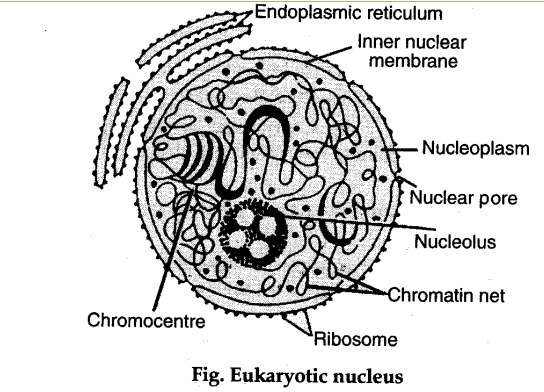
Describe the structure of nucleus and centrosome with the help of labelled diagram - CBSE Class ...
diagram of cell membrane labeled diagram of cell membrane labeled Interactive Eukaryotic Cell Model we have 9 Images about Interactive Eukaryotic Cell Model like Interactive Eukaryotic Cell Model, This micrograph shows the structure of a pancreatic acinar cell and the and also Interactive Eukaryotic Cell Model. Here you go: Interactive Eukaryotic Cell Model

tech_savvy_reynolds: Cell membrane diagram created with Inspiration
Cell: Structure and Functions (With Diagram) - Biology Discussion 1. Eukaryotes are sophisticated cells with a well defined nucleus and cell organelles. 2. The cells are comparatively larger in size (10-100 μm). 3. Unicellular to multicellular in nature and evolved ~1 billion years ago. 4. The cell membrane is semipermeable and flexible. 5.
Micro-organisms: Cells Flashcards | Easy Notecards
diagram of cell membrane labeled Cell Nucleus biologywise.com. nucleus. Labeling Exercise - Dr. Hunter Biology sites.google.com. cell membrane worksheet answers answer key function structure biology corner title concept map worksheets labeling blood briefencounters worksheeto. 1.3 Membrane Structure - BIOLOGY4IBDP biology4ibdp.weebly.com. membrane structure. Diagram Of A Cell ...
Membranes I | Biology | Visionlearning
PDF Human Cell Diagram, Parts, Pictures, Structure and Functions The cell membraneis the outer coating of the cell and contains the cytoplasm, substances within it and the organelle. It is a double-layered membrane composed of proteins and lipids. The lipid molecules on the outer and inner part (lipid bilayer) allow it to selectively transport substances in and out of the cell. Endoplasmic Reticulum

Nucleus, the commanding centre of the cell ~ Biology Exams 4 U
Basic Cell Membrane Label - Labelled diagram - Wordwall Integral Protein (channel), Peripheral Protein, Phosphate, Lipid, Hydrophilic, Hydrophobic, Glycoprotein.
Introduction To Cell Membrane Functions - Cell Diagram
Plasma Membrane Function, Structure & Diagram - Study.com The plasma membrane name comes from its ability to move with the cell in a flexible way, like plasma flowing, and its function in creating a barrier, or membrane to separate the cell from the ...

Cell Membrane Diagram Labeled : Functions and Diagram
CELL MEMBRANE LABEL Diagram | Quizlet Practice labeling the parts of the cell membrane Terms in this set (6) Channel Protein hole or tunnel that particles may pass through to go in / out of cell Marker protein identifies or labels the cell Receptor protein receives information Heads part of the phospholipid that loves water (hydrophili) - points to the most outside and inside of cell
/2000px-Plant_cell_structure_svg_vacuole.svg-58a886443df78c345bf8d009.png)
Vacuole Organelle
Animal Cells: Labelled Diagram, Definitions, and Structure The endoplasmic reticulum (s) are organelles that create a network of membranes that transport substances around the cell. They have phospholipid bilayers. There are two types of ER: the rough ER, and the smooth ER. The rough endoplasmic reticulum is rough because it has ribosomes (which is explained below) attached to it.

Animal And Plant Cells.
Plant Cells: Labelled Diagram, Definitions, and Structure Plastids and Chloroplasts. Plants make their own food through photosynthesis. Plant cells have plastids, which animal cells don't. Plastids are organelles used to make and store needed compounds. Chloroplasts are the most important of plastids. They convert light energy from the sun into sugar and oxygen. The most exposed parts of the plants ...

Animal Name Cell Membrane / Name: Animal Cell Coloring Sheet Cell Membrane (ligh brown ...
Labeled Plant Cell With Diagrams - Science Trends The parts of a plant cell include the cell wall, the cell membrane, the cytoskeleton or cytoplasm, the nucleus, the Golgi body, the mitochondria, the peroxisome's, the vacuoles, ribosomes, and the endoplasmic reticulum. Parts Of A Plant Cell The Cell Wall Let's start from the outside and work our way inwards.

Cell Membrane A Level Biology Functions - Cell Diagram
Cell Membrane (Plasma Membrane) - Genome.gov Definition. …. The cell membrane, also called the plasma membrane, is found in all cells and separates the interior of the cell from the outside environment. The cell membrane consists of a lipid bilayer that is semipermeable. The cell membrane regulates the transport of materials entering and exiting the cell.

Detailed Diagram Models Cell Membrane Stock Vector 376416385 - Shutterstock
Structure of Membrane in Cells (With Diagram) - Biology Discussion Cell walls are formed through apposition, i.e., the wall material is deposited by the protoplast on the plasma membrane. The first cell wall (Primary wall) is formed during the cell growth phase. When the cell elongation is stopped the secondary cell wall, formation starts (Fig. 2.13).

Generalized Plant Cell
Diagram of a cell membrane with labels - NIST Diagram of a cell membrane with labels | NIST IMAGES Diagram of a cell membrane with labels Appears In Biology in Reflectometry Essential Biological Functions Immune response, Cell metabolism, Neurotransmission, Photosynthesis, Cell adherence, Cell growth and differentiation Potential Commercial Applications

Cell Membrane - Organelles & Beyond

Membranes Organize Cellular Complexity | Biology teacher, Biology classroom, Teaching biology

Interactive cell diagram by Diann Caviness

Prokaryotic Cell
Post a Comment for "43 cell membrane diagram with labels"