45 ear anatomy without labels
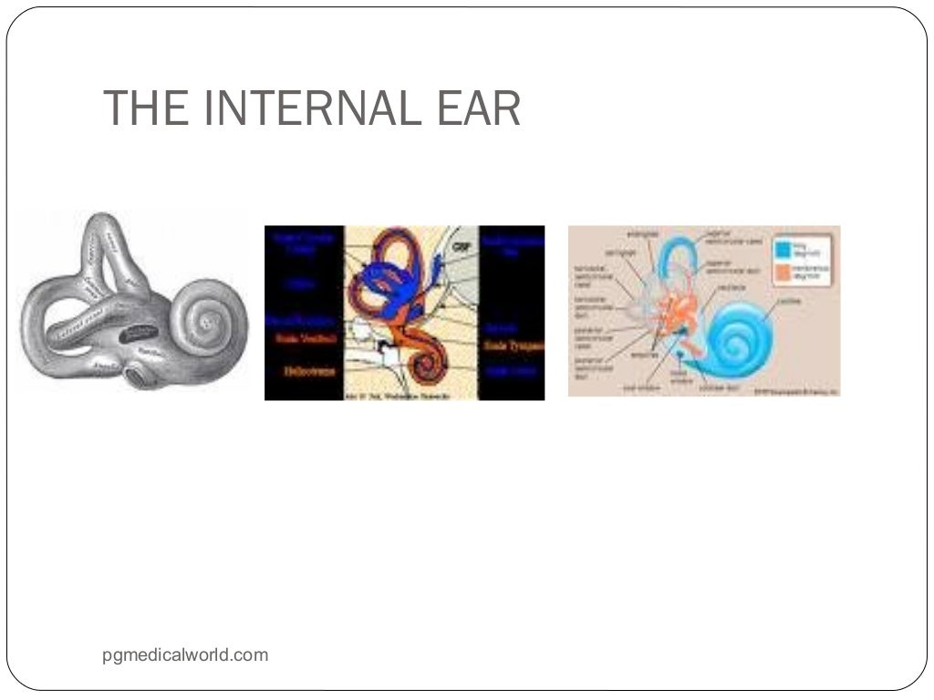
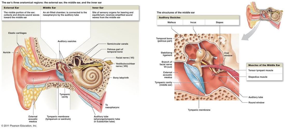
Ear Anatomy, Diagram & Pictures | Body Maps - Healthline A bony casing houses a complex system of membranous cells. The inner ear is called the labyrinth because of its complex shape. There are two main sections within the inner ear: the bony labyrinth ... Ear (Anatomy): Overview, Parts and Functions | Biology Dictionary The human ear picks up and interprets high-frequency vibrations of air, while the sound-sensing organs of aquatic animals are designed to pick up high-frequency vibrations in water. Most vertebrates have two ears: one on either side of the head. In some animals, including most mammals, the ear is also used for balance.
Human Body Parts Images Without Labels - Free Vector Download 2020 Human ear diagram with labels and label of anatomy labeling the ear purposegames nose diagram with label diagrams all labels human ear the ear diagram without labels anatomy human charts. Illustration Of Body Parts Labels It is certainly the most widely studied structure the world over. Human body parts images without labels. Download body ...

Ear anatomy without labels
How To Draw A Eye And Ear And Label Them All The ears top should be along the eyebrow line and the bottom should closely line up to the nose. Parts of the Ear. Eyes drawing - step 2. Label The Parts Of The Eye. Okay shade in the inside outer edge of the inner circle of the eye yes that is a lot of words. Select the correct label for each part of the ear. Tylenol® PM and High Blood Pressure | TYLENOL® You’re not alone. Nearly half of all US adults suffer from high blood pressure 1 (hypertension) and in one study 80% of patients with high blood pressure were unaware that NSAIDs can interfere with certain antihypertensives 2.The CDC also found that >1 in 3 adults don’t get enough sleep on a regular basis 3.. Know the facts Applied Sciences | Free Full-Text | A Novel Immersive Anatomy … Jun 03, 2022 · Immersive technologies are redefining ways of interacting with 3D objects and their environments. Moreover, efforts in blended learning have presented several advantages of incorporating educational technology into the learning space. The advances in educational technology have in turn helped to widen the choice of different pedagogies for improving …
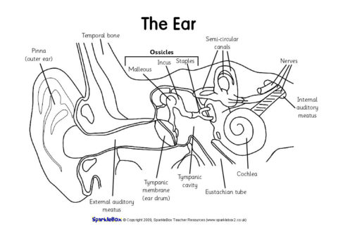
Ear anatomy without labels. Image result for ear structure without label | Ear diagram, Human ear ... Image result for ear structure without label. Find this Pin and more on • S C I E N C E • by Passant Ghonim. Human Ear Anatomy. Human Anatomy Picture. Human Ear Diagram. Interview Thank You Letter. Ear Structure. Label Templates. Study Notes. Human Ear Anatomy - Parts of Ear Structure, Diagram and Ear Problems The external (outer) ear consists of the auricle, external auditory canal, and eardrum (Figure 1 and 2). The auricle or pinna is a flap of elastic cartilage shaped like the flared end of a trumpet and covered by skin. The rim of the auricle is the helix; the inferior portion is the lobule. Ligaments and muscles attach the auricle to the head. Ear Anatomy - Outer Ear | McGovern Medical School The medical term for the outer ear is the auricle or pinna. The outer ear is made up of cartilage and skin. There are three different parts to the outer ear; the tragus, helix and the lobule. EAR CANAL The ear canal starts at the outer ear and ends at the ear drum. The canal is approximately an inch in length. Ear Diagram and Labeling Worksheet - Twinkl The first worksheet presents an ear with annotations showing the first letters of its key features. For example, a label marked 'P' links to the Pinna (outer ear). The second page shows an ear diagram without labels. The final page shows the labels linking to the beginning letters of each feature, but without the words list.
Atlas of Human Anatomy: Including Student Consult Interactive ... The gold standard of excellence for 25 years, Frank H. Netter, MD’s Atlas of Human Anatomy offers unsurpassed depictions of the human body in clear, brilliant detail – all from a clinician’s perspective. With its emphasis on anatomic relationships and clinically relevant views, Dr. Netter’s work provides a coherent, lasting visual vocabulary for understanding anatomy and how it … Human Ear Diagram - Bodytomy External Auditory Canal: External auditory canal or ear canal, is the channel from which the sound enters from the outside ear to the eardrum. Eardrum/Tympanic Membrane: It is the thin membrane located between the external ear canal and the middle ear. Cochlea: Cochlea is tiny conical structure situated in the inner ear that resembles a snail shell. It is responsible for converting sound vibrations into nerve signals that are sent to the brain. The Ear: Anatomy, Function, and Treatment - Verywell Health The middle ear (also known as the tympanum or tympanic cavity) is a complicated network of tunnels, chambers, openings, and canals mostly inside openings within the temporal bone on each side of the skull. The 2 largest chambers are called the middle ear space and mastoid. e-Anatomy: radiologic anatomy atlas of the human body - IMAIOS e-Anatomy is an award-winning interactive atlas of human anatomy. It is the most complete reference of human anatomy available on web, iPad, iPhone and android devices. Explore over 6,700 anatomic structures and more than 870,000 translated medical labels. Images in: CT, MRI, Radiographs, Anatomic diagrams and nuclear images. Available in 12 ...
Anatomy of the Ear - Geeky Medics Figure 1.Anatomy of the external ear. 4 Innervation of the auricle. The auricle has several sources of sensory innervation:. The superficial surface is supplied by the great auricular nerve and lesser occipital nerve, both of which are branches of the cervical plexus (C2 & C3), and the auriculotemporal branch of the mandibular nerve, which is a branch of the trigeminal nerve (cranial nerve V) Ear Anatomy without Labels, Digital Art - Shutterstock Ear Anatomy Without Labels Digital Art Stock Illustration 530108302 Edit Download for free See more Popularity score High Usage score High usage Superstar Shutterstock customers love this asset! Item ID: 530108302 Ear Anatomy without Labels, Digital Art Formats 8976 × 6201 pixels • 29.9 × 20.7 in • DPI 300 • JPG Picture of the Ear: Ear Conditions and Treatments - WebMD The ear has external, middle, and inner portions. The outer ear is called the pinna and is made of ridged cartilage covered by skin. Sound funnels through the pinna into the external auditory... flower diagram without labels 10 Best Images Of Label Ear Diagram Worksheet - Blank Rock Cycle . diagram cycle rock blank label worksheet ear key worksheeto via. Nature, Cultural, And Travel Photography Blog: 1/1/15 - 2/1/15 pk-photography.blogspot.com. zealand natural beauty birdy landscapes labels official posted
Pin by elizabeth wright on Free time | Human skull anatomy, Skull anatomy, Skull
Blank ear diagrams and quizzes: The fastest way to learn - Kenhub That's why labeling the ear is an effective way to begin your revision. It helps you to memorize the names and their locations, which in turn will aid you to remember their functions. Below, you can download both the blank ear diagram to make some notes, and then try labeling the ear using the unlabeled ear diagram. Good luck!
Label the Ear Worksheets (SB6635) - SparkleBox
Parts of the Ear Labelled Diagram Activity - Twinkl The first worksheet presents an ear with annotations showing the first letters of its key features. For example, a label marked 'P' links to the Pinna (outer ear). The second page shows an ear diagram without labels. The final page shows the labels linking to the beginning letters of each feature, but without the words list.
(184).jpg)
Anatomy Of The Ear - ProProfs Quiz
Anatomy coloring books: How to use & free PDF | Kenhub Sep 30, 2021 · Generally, an anatomy coloring book will divide subject matter into sections, with each section containing many topics. For each topic you will find black and white anatomical drawings, often accompanied by labels, related text and terminology. Tired of keeping track of so many study materials?
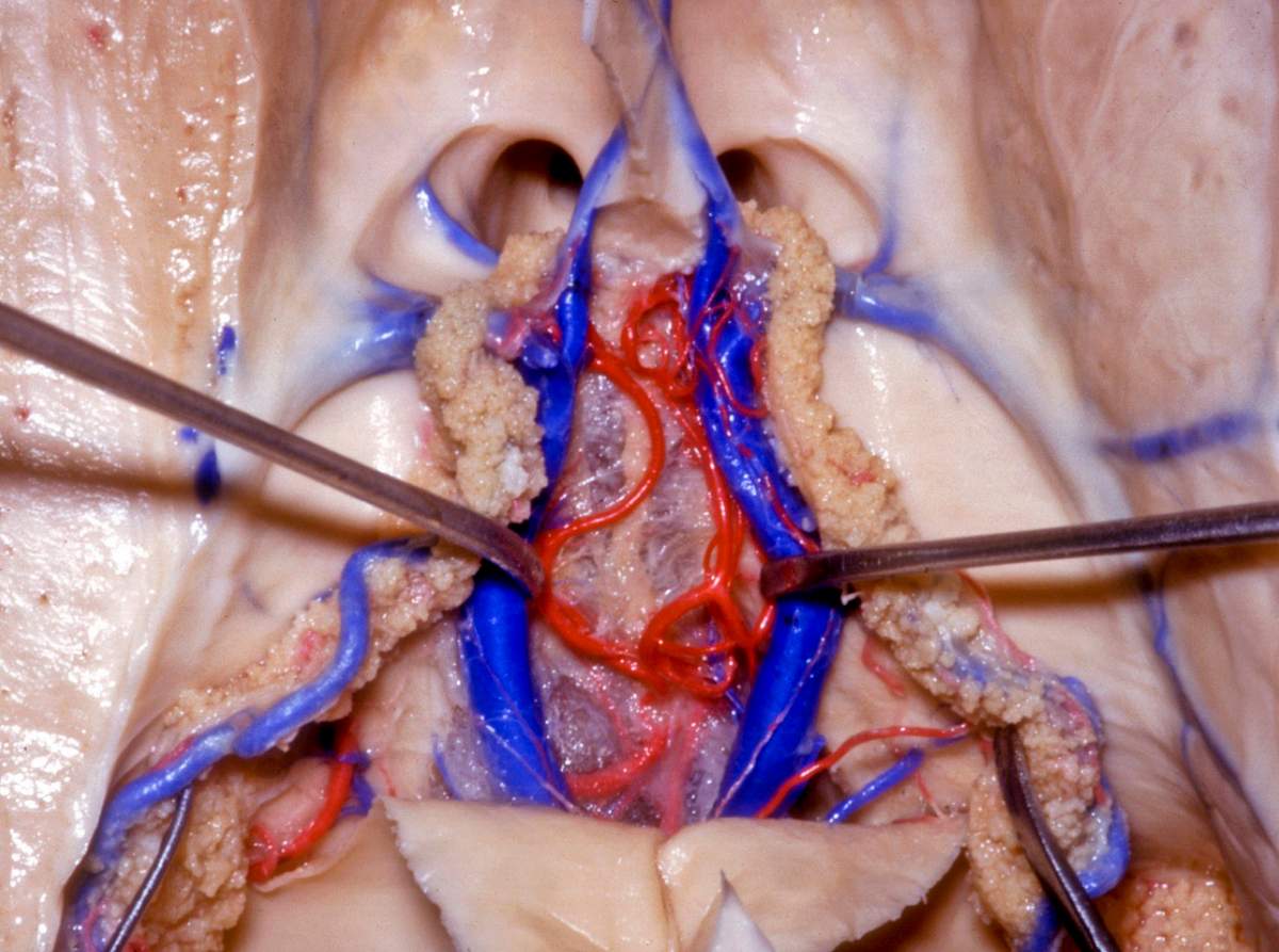
Velum Interpositum and Choroidal Vessels | Neuroanatomy | The Neurosurgical Atlas, by Aaron ...
enchantedlearning.com Moved Permanently. The document has moved here.
Anatomy of ear
TeachMeAnatomy - Making Anatomy Simple Containing over 1000 vibrant, full-colour images, TeachMeAnatomy is a comprehensive anatomy encyclopaedia presented in a visually-appealing, easy-to-read format.. Created by a team of doctors and medical students, each topic combines anatomical knowledge with high-yield clinical pearls, seamlessly bridging the gap between scholarly learning and ...

31 Label The Ear Diagram - Labels Database 2020
The Human Ear - Structure, Functions and its Parts - BYJUS There are three ear ossicles in the human ear: Malleus: A hammer-shaped part that is attached to the tympanic membrane through the handle and incus through the head. It is the largest ear ossicle. Incus: An anvil-shaped ear ossicle connected with the stapes. Stapes: It is the smallest ossicle and also the smallest bone in the human body. Inner Ear

Category:SVG all portions of the human ear - Wikimedia Commons
Cool Label The Ear Anatomy Diagram Label the ear anatomy diagram. Below you can download both the blank ear diagram to make some notes and then try labeling the ear using the unlabeled ear diagram. Incorrect answers will be marked in red. Memorizing the parts of the ear isnt difficult. Start studying Ear Anatomy Labeling Practice SPECIAL SENSES. The Ear - Science Quiz.

Oral Cavity Gallery - Medical Information Illustrated
ear diagram without labels ear diagram without labels Label Parts of the Human Ear we have 9 Images about Label Parts of the Human Ear like Ear Labeling Quiz - Human Anatomy, Label Parts of the Human Ear and also 10 Best Images of Label Ear Diagram Worksheet - Blank Rock Cycle. Here it is: Label Parts Of The Human Ear academic.udayton.edu
Ear | ClipArt ETC
(PDF) LIBRO PARA COLOREAR NETTER - Academia.edu Enter the email address you signed up with and we'll email you a reset link.

This excellent ear diagram labels all the important parts of the human ear system. The labeled ...
Petrous bone CT: normal anatomy| e-Anatomy - IMAIOS Sep 13, 2021 · Anatomy of the temporal bone: how to view the anatomical labels. This module is a comprehensive and affordable learning tool for residents and medical students and specially for neuroradiologists and otolaryngologists. It provides images in the axial and coronal planes, allowing the user to review and learn anatomy interactively.

Human ear Royalty Free Vector Image - VectorStock
Cefdinir Antibiotic Side Effects, Uses (Strep, Middle Ear) & Dosage Cefdinir is an antibiotic in the cephalosporin drug class prescribed to treat infections, for example, middle ear, tonsillitis, strep throat, bronchitis, and sinusitis. Common side effects are nausea, abdominal pain, loose stools, and vaginitis. Dosage and pregnancy and breastfeeding safety information are included.

Susan Price's 'Nennius' Blog: I Want To Write!
Ear Anatomy: Understanding the Outer, Middle, and Inner Parts of the Ear In this section, we describe the anatomy of the ear in simple terms. External Ear Anatomy (Auricle or Pinna) The outer ear auricle or external ear is composed of all of the parts of the ear outside the skull. It is also sometimes referred to as the auricle or the pinna. Although the outer ear is the least important part of the ear's hearing function, it provides the necessary structure and protection.
13 Best Images of Frog Anatomy Labeling Worksheet - Frog Dissection Diagram Labeled, Labeled ...
Health Encyclopedia The ear is the organ of hearing and balance. The parts of the ear include: Pinna or auricle. This is the outside part of the ear. External auditory canal or tube. This is the tube that connects the outer ear to the inside or middle ear. Tympanic membrane (eardrum). The tympanic membrane divides the external ear from the middle ear.
Chap 10 Anatomy
Label Parts of the Human Ear - University of Dayton Label Parts of the Human Ear. Select One Auditory Canal Cochlea Cochlear Nerve Eustachian Tube Incus Malleus Oval Window Pinna Round Window Semicircular Canals Stapes Tympanic Membrane Vestibular Nerve. Select One Auditory Canal Cochlea Cochlear Nerve Eustachian Tube Incus Malleus Oval Window Pinna Round Window Semicircular Canals Stapes Tympanic ...
Post a Comment for "45 ear anatomy without labels"